Gallery
GDV method
Gas Discharge Visualization Technique (GDV) is computer registration and analysis of gas discharge glow (GDV-images) of any biological objects placed in a high intensity electromagnetic field.
When more than 70 years ago the married couple Kirlians started to study high-frequency photography it was regarded just as a scientific experiment.
Modern technologies and the rhythm of our life bringing hazards to health and leaving less and less time to take care of it have changed this view.
Instead of emergency medicine preventive medicine has come into picture. Fast methods of diagnostics appeared uniting electrophysiological and clinical-anatomical characteristics of body. Among them are electroencephalogram (EEG), electrocardiogram, (ECG) and others.
Development of technologies made it possible to elaborate a bioelectrographic method of complex express diagnostics of human body and evaluation of factors and treatments that can influence it.
On the basis of Kirlian effect a team of scientists under the guidance of Prof. Korotkov developed the GDV Technique and created a device to produce gas discharge images of objects (human fingers, in particular).
The GDV Technique is patented, the hardware passed technical, toxicological and clinical tests; it is registered in the public register of medical equipment in the Ministry of Health of the Russian Federation.
During 10 years the GDV Technique has won the recognition of many specialists and together with other electrographic methods is used in medicine, professional sports and fitness, sanatoria and health resorts, the beauty business, various fields of psychology and psychophysiology, and also in basic and applied research.
The GDV Technique was developed in Saint Petersburg by Professor K. G. Korotkov on the basis of Kirlian effect.
Subjects’ studies by GDV Technique involve connecting an object on the glass electrode to the electrical circuit of the device that forms impulses of a high intensity electromagnetic field (duration 10 µs, frequency 1024 Hz). As a result, a sequence of gas discharges is formed within a preset exposure time. Gas discharge developing in the impulse acts as an amplifier of super-low emission processes taking place on the surface of the subject; at the same time the surface distribution of discharge channels depends on the topography of electro-physical characteristics of the subject.

GDV-image is a complex 2-D figure with each pixel characterized by its brightness coded by integer in the range of 0 ("black") to 255 ("white"). Geometrical parameters of GDV-images (e.g., the area defined as a sum of pixels exceeding the specified brightness threshold; fractality coefficient defined as relation of the length of the image perimeter to its average radius multiplied by 2Pi, the breadth of streamers) contain the information about the object’s characteristics. For example, with an increase of ion concentration in liquid the glow increases while the streamer breadth decreases. To include such data into the structure of a complex biophysical experiment quantitative processing of the obtained images is required.

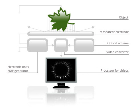
The principle of image formation is as follows. Between the object under study and the transparent electrode on which the object is placed the voltage impulses are applied from an electromagnetic field generator. For this purpose there is a transparent current conductive coating layer applied on the other side of the glass electrode. With a high intensity field in the gas medium of the contact space of the object and electrode glass an avalanche or sliding gas discharge is formed whose characteristics are determined by the object’s properties. The optical system and CCD matrix transform the discharge glow into video signals that are registered as single frames (GDV-images) or AVI-files in the memory unit connected with the processor that carries out the video frames processing. The processor is a specialized software complex that enables to calculate a whole number of parameters and to produce certain diagnostic conclusions on their basis.
User login
Last articles


